
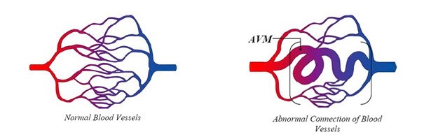
Normally, arteries carry blood containing oxygen from the heart to the brain, and veins carry blood with less oxygen away from the brain and back to the heart. When an arteriovenous malformation (AVM) occurs, a tangle of blood vessels in the brain or on its surface bypasses normal brain tissue and directly diverts blood from the arteries to the veins.
As the name suggests, vascular malformations of the brain is an umbrella term for at least six conditions in which blood vessels of the brain are affected. Such malformations are classified into several types in which the symptoms, severity, and causes vary. These types of VMB are: (1) arteriovenous malformations (AVM), abnormal arteries and veins; (2) cavernous malformations (CM), enlarged blood-filled spaces; (3) venous angiomas (VA), abnormal veins; (4) telangiectasias (TA), enlarged capillary-sized vessels; (5) vein of Galen malformations (VGM); and (6) mixed malformations (MM).
The symptoms you might experience depend on the type of vascular malformation you have, its size and where it is located in your head. Most of the time, vascular malformations cause no symptoms at all.
The tests and investigations you have might show up other factors which could affect your risk of experiencing any of the symptoms associated with vascular malformations. For example, their exact location and the routes veins take from them.
Most AVMs are detected with either a computed tomography (CT) brain scan or a magnetic resonance imaging (MRI) brain scan.
A doctor may also perform a cerebral angiogram. This test involves inserting a catheter (small tube) through an artery in the leg (groin). Then it’s guided into each of the vessels in the neck going to the brain, and a contrast material (dye) is injected and pictures are taken of all the blood vessels in the brain. For any type of treatment involving an AVM, an angiogram may be needed to better identify the type of AVM.
AVM Embolization
A small plastic tube, or catheter, is inserted through the groin and is guided up to the brain vessels and into the AVM. A glue, non-reactive liquid, is injected into the AVM. The adhesive material hardens as it is injected and blocks the blood flow through the AVM. If the AVM is larger in size, AVM embolization must be done in stages to ensure that all of the AVM is blocked off. AVM embolization is performed with the patient under general anesthesia.
Doctors often recommend AVM embolization prior to other AVM treatment options; embolization can reduce the size of the AVM and make it more responsive to radiation or suitable for surgical removal. AVM embolization often does not completely block the hemorrhage and must then be combined with other treatments after the AVM has been reduced in size. Once the AVM’s blood flow is reduced through embolization, surgical removal is faster and more successful.
AVM Radiation Treatment
For smaller AVMs, x-rays are used to thicken the blood vessels of the AVM and cut off the hemorrhage. The AVM often requires about 2 years to fully clot, and the risk of bleeding persists until the AVM has been completely eliminated.
AVM Surgery (resection)
The neurosurgeon opens the patient’s skull in order to see the AVM and clip the blood vessels that feed into it, in order to remove the AVM from the surrounding brain tissue. The arteries supplying the surrounding brain tissue are left intact; only scar tissue is removed with the AVM.
Surgically removing the AVM cures the patient immediately. AVMs do not grow back, and so the risk of bleeding is eliminated. However, only some AVMs may be safe for operation, depending on the size and location within the brain. AVM surgery is performed with the patient under general anesthesia.
What is head injury?A traumatic brain injury, also referred to an acquired brain injury, occurs when someone suffers a sudden…
read more
What is Brain Aneurysm?Brain aneurysm is an abnormal bulge in the brain's blood vessel. When it leaks or ruptures, it…
read more
What is a brain arteriovenous malformation ?Normally, arteries carry blood containing oxygen from the heart to the brain, and veins…
read more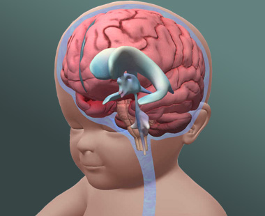
What is Hydrocephalus?Hydrocephalus is commonly referred to as "water on the brain." The so-called "water" is actually cerebrospinal fluid (CSF),…
read more