All posts by admin

Brain aneurysm

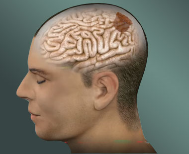
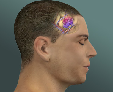
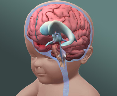
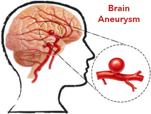
What is Brain Aneurysm?

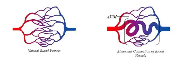
Brain aneurysm is an abnormal bulge in the brain’s blood vessel. When it leaks or ruptures, it causes bleeding into the brain, otherwise known as hemorrhagic stroke. A type of hemorrhagic stroke called subarachnoid hemorrhage occurs in the space in between the brain and the tissues covering it. This is the type of ruptured aneurysm that requires emergency treatment as it is life threatening.

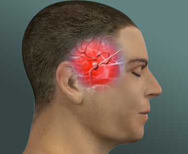
What are the Signs and Symptoms of Brain Aneurysm or Cerebral Aneurysm?

A ruptured aneurysm always exhibits the following common symptoms:

  • Headache that is extremely severe and sudden
  • Vomiting and nausea
  • Stiff neck
  • Double vision (blurred)
  • Light sensitivity
  • Seizure
  • An eyelid that drops
  • Losing consciousness
  • Being confused.

Leaking Brain Aneurysm

A small amount of blood can leak as a result of aneurysm leak. Severe headache that is sudden can only be caused by the leaking (sentinel blood).A leaking is always followed by a more serious rupture.

Un-Ruptured Brain Aneurysm

If the un-ruptured aneurysm is small it may not cause any symptoms at all. Nerves and tissues on the brain can be pressed by un-ruptured aneurysm that is large and it causes:

  • Pain behind and above the eye
  • The pupil is normally dilated
  • Double vision or change in visibility
  • One side of the face will be weak and numb or paralyzed
  • The eyelid will droop.
  • Development of a sudden headache that is extremely severe
  • must prompt you to seek medical attention immediately.


What are the causes of brain Aneurysm?

There are many conditions and factors that can lead to the formation of brain aneurysms.
 
The most common are smoking and uncontrolled high blood pressure. There are also genetic conditions including connective tissue disorders and polycystic kidney disease that can lead to aneurysm formation.


How is brain aneurysm diagnosed?

The majority of brain aneurysms never cause health problems; because of that, many go undiagnosed. Brain aneurysms typically are discovered only after a rupture, when the unruptured aneurysm is causing head pain, or when someone is undergoing tests for another condition. There are four main tests that can detect a brain aneurysm.

  • Angiography (also referred to as a cerebral angiogram) is a test in which dye is injected in the body. As the dye travels through veins and arteries, x-rays are taken and analyzed to determine the site of the aneurysm.
  • A computed tomography (CT) scan provides a detailed x-ray of the head that shows images of the brain and skull layer by layer.
  • A Magnetic resonance imaging (MRI) is another way to capture an image of the brain in detail.
  • Cerebrospinal fluid analysis is a test in which fluid is extracted from the space between the spinal cord and surrounding membrane and is tested for signs of bleeding or hemorrhage.

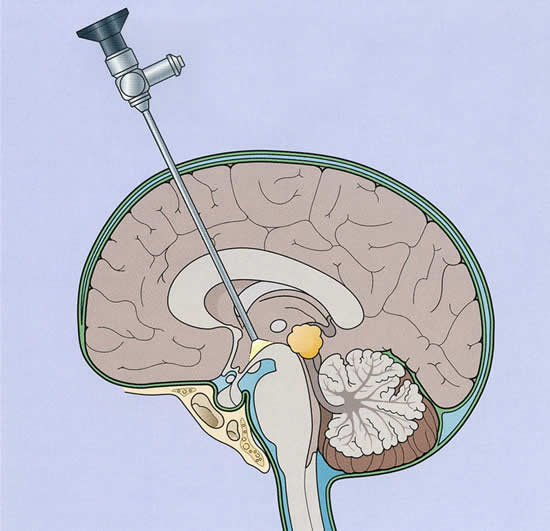
How is brain aneurysm treated?

The two common treatment methods for ruptured brain aneurysm are surgical clipping and endovascular coiling.

Surgical clipping involves a procedure of closing off an aneurysm. The doctor places a metal clip on the blood vessel that feeds the aneurysm to stop blood from flowing through it.

Endovascular coiling is less invasive. A surgeon inserts a catheter into the artery from the groin. Through this catheter, a wire is inserted to coil up inside the aneurysm to disrupt the blood flow.


Brain tumors

What is a brain tumor?

A brain tumor is a collection, or mass, of abnormal cells in your brain. Your skull, which encloses your brain, is very rigid. Any growth inside such a restricted space can cause problems. Brain tumors can be cancerous (malignant) or noncancerous (benign).

What are the causes of a brain tumor?

The exact cause of brain tumor is still not known, but studies suggest that certain risk factors might trigger brain tumor. Children receiving radiation are more prone in developing brain tumor as adults. A rare genetic condition called Li-Fraumeni syndrome or neurofibromatosis. People who are over the age of 65 are also at a higher risk of developing brain tumor. Cancerous brain tumor can lead to brain cancer and spread to the central nervous system and other part of the brain as well.

What are the symptoms of brain tumor?

Brain tumor symptoms can vary according to tumor type and location. There are times a person may have no symptoms when their brain tumor is discovered
• Recurrent headaches
• Issues with vision
• Seizures
• Changes in personality
• Short-term memory loss
• Poor coordination
• Difficulty speaking or comprehending
Whatever symptoms you have, discuss them fully with your physician so everyone has the most accurate information.

How is brain tumor diagnosed? 

Neurologic exam: Your doctor checks your vision, hearing, alertness, muscle strength, coordination, and reflexes.
MRI: A large machine with a strong magnet linked to a computer is used to make detailed pictures of areas inside your head.

CT scan: An x-ray machine linked to a computer takes a series of detailed pictures of your head.

Electroencephalography (EEG): An EEG is a noninvasive test in which electrodes are attached to the outside of a person’s head to measure electrical activity of the brain. It is used to monitor for possible seizures

Other tests will help determine specific information about the tumor which will be important to make a prognosis and treatment plan. Tests may include:

Blood, urine, and tissue tests—May be used to detect tumor markers, specific proteins that are elevated in the presence of cancer. Blood and tissue tests can be used to look for chromosome changes or inherited disorders associated with certain types of brain tumors.

Lumbar puncture —A needle is used to collect a sample of cerebrospinal fluid that surrounds and protects the brain and spinal cord. The test can determine if cancer cells are present in the fluid.

Biopsy: A biopsy is the removal of a small piece of suspicious tissue for examination under a microscope. Biopsies can also help determine if the tumor may have a higher rate of metastasis, meaning they are more likely to spread elsewhere in the brain. In some cases, the location of the tumor makes it difficult or impossible to get a biopsy. Imaging tests may be able to determine enough information so that a biopsy is not needed.

How is brain tumor treated?

Treatment for brain tumors depends on a number of factors including the type, location and size of the tumor as well as the patient’s age and general health. Treatment methods and schedules differ for children and adults. The most common treatment for brain tumors is surgery. Surgery can be combined with other treatments, such as radiation therapy and chemotherapy. Physical therapy, occupational therapy, and speech therapy can help recover after neurosurgery.

Vascular malformation

What is a brain arteriovenous malformation ?

Normally, arteries carry blood containing oxygen from the heart to the brain, and veins carry blood with less oxygen away from the brain and back to the heart. When an arteriovenous malformation (AVM) occurs, a tangle of blood vessels in the brain or on its surface bypasses normal brain tissue and directly diverts blood from the arteries to the veins.

As the name suggests, vascular malformations of the brain is an umbrella term for at least six conditions in which blood vessels of the brain are affected. Such malformations are classified into several types in which the symptoms, severity, and causes vary. These types of VMB are: (1) arteriovenous malformations (AVM), abnormal arteries and veins; (2) cavernous malformations (CM), enlarged blood-filled spaces; (3) venous angiomas (VA), abnormal veins; (4) telangiectasias (TA), enlarged capillary-sized vessels; (5) vein of Galen malformations (VGM); and (6) mixed malformations (MM).

What are the symptoms of vascular malformations of the brain?

The symptoms you might experience depend on the type of vascular malformation you have, its size and where it is located in your head. Most of the time, vascular malformations cause no symptoms at all.

The tests and investigations you have might show up other factors which could affect your risk of experiencing any of the symptoms associated with vascular malformations. For example, their exact location and the routes veins take from them.

  • Strokes- Strokes affecting people with vascular malformations are usually due to bleeding in the brain (a haemorrhage).
  • Epilepsy-For people with an AVM or cavernous malformation of the brain, in any given year there is a 1 in 100 (1%) risk of developing epilepsy for the first time.
  • Headache- Headaches are a common problem for a large number of people and it is sometimes difficult to establish whether they are related to a vascular malformation or have a different cause.People with vascular malformations can have headaches that are similar to migraines and might respond to migraine treatments.
  • Visual problems -Visual problems such as blurred vision, double vision or a loss of vision, particularly affecting one eye, can be a symptom of stroke.
  • Noises in the head-An AVF of the dura can cause a noise in your head due to the blood flowing through it. The noise is called a bruit. A doctor can hear it using a stethoscope. Occasionally, you might be able to hear the noise yourself. Some people hear it as a distinctive type of ringing or whooshing sound that beats in time with their pulse. This is called pulsatile tinnitus.
  • Memory problems-If you have had a brain haemorrhage, you might be left with damage to your brain which can cause problems with your short-term memory, attention and concentration.Even without having had a haemorrhage, people with AVMs sometimes report having problems with their memory. This is possibly due to changes in the flow of blood through their brain.
  • Anxiety and depression
  • Other symptoms-Other symptoms of vascular malformations of the brain might include speech problems, weakness in the arm or leg, balance problems, and enlarged blood vessels on the scalp.

How are AVMs diagnosed?

Most AVMs are detected with either a computed tomography (CT) brain scan or a magnetic resonance imaging (MRI) brain scan.
A doctor may also perform a cerebral angiogram. This test involves inserting a catheter (small tube) through an artery in the leg (groin). Then it’s guided into each of the vessels in the neck going to the brain, and a contrast material (dye) is injected and pictures are taken of all the blood vessels in the brain. For any type of treatment involving an AVM, an angiogram may be needed to better identify the type of AVM.

Common brain AVM treatments include:

AVM Embolization

A small plastic tube, or catheter, is inserted through the groin and is guided up to the brain vessels and into the AVM. A glue, non-reactive liquid, is injected into the AVM. The adhesive material hardens as it is injected and blocks the blood flow through the AVM. If the AVM is larger in size, AVM embolization must be done in stages to ensure that all of the AVM is blocked off. AVM embolization is performed with the patient under general anesthesia.

Doctors often recommend AVM embolization prior to other AVM treatment options; embolization can reduce the size of the AVM and make it more responsive to radiation or suitable for surgical removal. AVM embolization often does not completely block the hemorrhage and must then be combined with other treatments after the AVM has been reduced in size. Once the AVM’s blood flow is reduced through embolization, surgical removal is faster and more successful.

AVM Radiation Treatment

For smaller AVMs, x-rays are used to thicken the blood vessels of the AVM and cut off the hemorrhage. The AVM often requires about 2 years to fully clot, and the risk of bleeding persists until the AVM has been completely eliminated.

AVM Surgery (resection)
The neurosurgeon opens the patient’s skull in order to see the AVM and clip the blood vessels that feed into it, in order to remove the AVM from the surrounding brain tissue. The arteries supplying the surrounding brain tissue are left intact; only scar tissue is removed with the AVM.

Surgically removing the AVM cures the patient immediately. AVMs do not grow back, and so the risk of bleeding is eliminated. However, only some AVMs may be safe for operation, depending on the size and location within the brain. AVM surgery is performed with the patient under general anesthesia.

Hydrocephalus (Endoscopic surgery)

What is Hydrocephalus?

Hydrocephalus is commonly referred to as “water on the brain.” The so-called “water” is actually cerebrospinal fluid (CSF), a clear liquid that looks like water and is produced in the 4 ventricles (cavities) of the brain, connected by narrow pathways.

What are the causes of hydrocephalus?

Hydrocephalus is caused by an imbalance between how much cerebrospinal fluid is produced and how much is absorbed into the bloodstream.

What are the symptoms of hydrocephalus?

The main symptoms of hydrocephalus are

  • headache
  • nausea (feeling sick)
  • vomiting (being sick)
  • confusion, and
  • problems with vision (for example, blurred or double vision).

Adults with hydrocephalus might also experience

  • balance problems
  • poor coordination
  • shuffling when they walk, and
  • urinary incontinence (a loss of control passing urine).
  • People would not necessarily experience all of these symptoms, but they would usually experience more than one of them.

In cases of normal pressure hydrocephalus (NPH), the build-up of CSF can be gradual and the symptoms of hydrocephalus might develop slowly over long periods of time (months and years rather than weeks).

Tests and investigations

The key test for hydrocephalus is a brain scan. This might be a CT (computerised tomography) scan or an MRI (magnetic resonance imaging) scan. The scan results can show signs of a build-up of CSF and increased pressure in the brain.

How is hydrocephalus treated?

There are no effective medicines for hydrocephalus. Most children require surgery. The goal is to lessen the pressure in the brain by providing another pathway for CSF to be drained and absorbed away from the brain.

There are two types of surgery for hydrocephalus:

Shunt surgery

The key treatment for hydrocephalus is a shunt. A shunt is a thin tube implanted in the brain to drain away the excess CSF to another part of the body (often the abdominal cavity, the space around the bowel) where it can be absorbed into the bloodstream.

Endoscopic third ventriculostomy (ETV).


Some children with non-communicating hydrocephalus can have surgery called an endoscopic third ventriculostomy (ETV). During an ETV, the surgeon makes an opening in the floor of the ventricle at the base of the brain. The CSF is then no longer blocked inside the ventricle. Now it can flow in and around the brain as it should.
This means that the child will not need a shunt, but instead will rely on the opening made by the surgeon during surgery.